
Diagnostic Imaging
What is Diagnostic Imaging?
Diagnostic imaging is a group of medical tests that create detailed pictures of the inside of your body. In orthopaedics, it helps your surgeon understand what may be causing pain, stiffness, swelling, weakness, or reduced movement. These images show bones, joints, tendons, ligaments, cartilage, and soft tissues so that your doctor can make an accurate diagnosis and design the right treatment plan. Imaging is often one of the first steps in assessing injuries or long-standing symptoms when a physical examination alone cannot give the full picture. It is a safe, commonly used tool that supports decision-making in both non-surgical and surgical care.
Imaging does not replace a clinical examination; instead, it adds extra clarity. Many orthopaedic conditions appear similar on physical exam, and imaging helps confirm or rule out fractures, arthritis, tendon damage, ligament tears, bursitis, or joint instability. Through these detailed pictures, your surgeon can track the severity of the problem, understand its impact on your movement, and determine the best path to recovery.

Who is Suitable for Diagnostic Imaging?
Diagnostic imaging is suitable for most patients experiencing musculoskeletal symptoms. It is used for adults, teenagers, elderly patients, and even children when necessary. Your surgeon may recommend imaging if you have:
- Persistent pain: Ongoing discomfort in the shoulder, elbow, wrist, hip, knee, ankle, foot, neck, or back
- Recent injuries: Trauma from falls, sports, work incidents, or motor vehicle accidents
- Reduced function: Difficulty walking, lifting, gripping, bending, or performing daily tasks
- Unclear symptoms: Swelling, numbness, locking, catching, instability, or unexplained weakness
- Monitoring of chronic conditions: Arthritis, tendon degeneration, or long-term joint wear
- Planning treatment: To guide injection therapy, physiotherapy, or surgery if required
Diagnostic imaging is generally safe for most people. Some imaging types, particularly those involving radiation, are used carefully during pregnancy unless urgent. Your surgeon will guide you to the safest and most appropriate test for your situation.

Benefits of Diagnostic Imaging
Diagnostic imaging plays a central role in high-quality musculoskeletal care. Its advantages include:
- Accurate diagnosis: Clear images help your surgeon identify the exact cause of your symptoms
- Better treatment decisions: Imaging ensures you receive the most effective and targeted care
- Early detection: Problems such as small fractures or early arthritis can be found before they worsen
- Monitoring progress: Imaging can assess how well treatments like physiotherapy or injections are working
- Avoiding unnecessary surgery: If imaging shows that non-surgical treatment is suitable, surgery may not be needed
- Safety and precision: Imaging can guide procedures such as injections, making them more comfortable and accurate
Overall, imaging supports your recovery by providing reliable information that improves treatment planning and outcomes.
Types of Diagnostic Imaging
Orthopaedic surgeons use several types of imaging depending on what needs to be assessed. Each method provides different information.
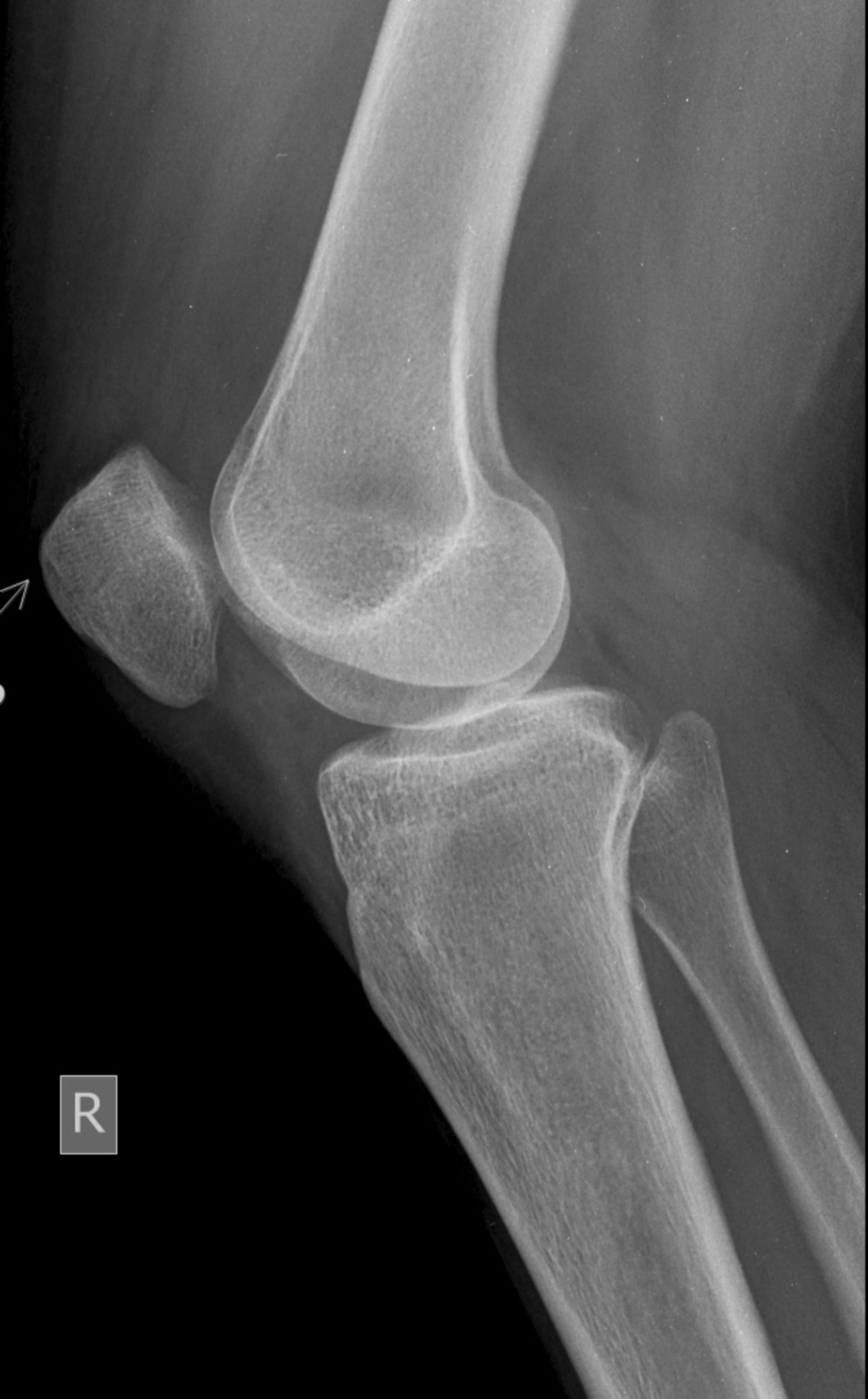
- X-ray: A quick, simple test that uses low-dose radiation to show bones and joints. They help diagnose fractures, arthritis, joint alignment issues, bone spurs, and long-term wear and tear. X-rays do not show soft tissues well, but provide essential baseline information.
- Ultrasound: Ultrasound uses sound waves to show tendons, ligaments, muscles, and soft tissues in real time. It is very useful for diagnosing tendon tears, bursitis, inflammation, and fluid collections. It can also guide injections with high accuracy.
- MRI scan: MRI creates highly detailed images of soft tissues, cartilage, ligaments, nerves, and bone marrow without radiation. It is particularly helpful for diagnosing meniscus tears, rotator cuff injuries, ligament injuries, cartilage damage, and early joint disease.
- CT scan: CT provides cross-sectional images of bones and joints. It is helpful for complex fractures, bone deformity, joint reconstruction planning, and conditions requiring high-resolution bone structure.
- Bone scan: A bone scan highlights areas of abnormal bone activity. It can detect stress fractures, infection, bone tumours, or widespread joint inflammation.
- DEXA scan: DEXA measures bone density and is often used to assess the risk of osteoporosis or fractures.
Your surgeon will select only the necessary tests, avoiding unnecessary radiation or cost.
Preparation Needed Before a Diagnostic Imaging
Preparing for diagnostic imaging is usually simple and depends on the type of test your surgeon has recommended. Most patients do not need to make major changes before their appointment. Your doctor or imaging clinic will give instructions specific to your scan, but there are general steps that can help you feel comfortable and ready.
- Wear loose clothing that does not restrict movement, as some scans require you to lie still or change positions.
- Remove jewellery, belts, and other metal accessories, as they may interfere with certain scans, particularly X-rays, CT scans, and MRI scans. Some imaging tests require no food or drink for a short time beforehand, such as CT scans with contrast dye, but most orthopaedic tests do not require fasting.
- Inform your surgeon and the clinic if you are pregnant, breastfeeding, or have metal implants, pacemakers, or medical devices, as these may influence the test type or safety precautions.
- Bring previous scan results if you have them, as these help the radiologist compare changes over time. Arrive a little early so that paperwork and preparation can be completed without stress.
- If you have concerns, anxiety, or claustrophobia (relevant for MRI), let our clinic know beforehand so they can offer support or options to help you remain calm.
What Happens During a Diagnostic Imaging?
The process during the imaging appointment depends on the scan type, but most tests are painless, straightforward, and completed within minutes.
- A trained radiographer or sonographer will guide you through each step and ensure you feel safe and comfortable. You may be asked to change into a gown or reposition yourself several times for accurate imaging.
- For X-rays, you will stand, sit, or lie down for a few seconds while the image is taken.
- For ultrasound, a small amount of gel is applied to the skin, and a handheld probe is moved across the area to capture images in real time.
- MRI scans require you to lie on a table that slides into a tube-shaped machine; the test is painless but can be noisy, and ear protection is always provided.
- For CT scans, the machine moves around the body to take rapid cross-sectional images. If contrast dye is needed, it may be given through a small cannula to help highlight structures.
- Bone scans involve a small injection of a tracer, followed by imaging after a short wait. Throughout each test, you will be asked to stay still to ensure the images are clear and accurate.
The radiographer monitors you closely, communicates each step, and ensures your comfort. A specialist radiologist reviews the test results and then sends them to your surgeon, who will explain the findings to you.
What to Expect After a Diagnostic Imaging?
Most patients can return to normal activities immediately after diagnostic imaging. There is usually no recovery time needed unless contrast dye or certain injections are involved. After X-rays, MRI, CT scans, or ultrasound, you can go home or back to work straight away. If contrast dye was used, you may be asked to drink extra water to help your body flush it out. Very occasionally, mild side effects such as a headache or brief warmth can occur, but they usually settle quickly.
Your surgeon will receive the radiology report and images electronically. In most cases, you do not have to collect anything from the imaging clinic. At your next appointment, your surgeon will explain the results, what they mean, and how they affect your treatment plan. Imaging can guide decisions about physiotherapy, injections, medication, or surgery, if needed. If your symptoms change or worsen after the scan, let your doctor know, as additional imaging or treatment adjustments may be required.
Remember that imaging alone does not treat the condition; instead, it helps your surgeon plan the most effective care. Many patients feel reassured once they understand the cause of their symptoms and have a clear path to recovery.
Diagnostic Imaging Prognosis
The prognosis after diagnostic imaging is excellent, as the test itself has no long-term impact on your health. Instead, its value lies in improving the accuracy and effectiveness of your diagnosis and treatment. When imaging clearly shows the structure and condition of bones, joints, and soft tissues, your surgeon can identify early problems, confirm suspected injuries, or rule out more serious concerns. This leads to better-targeted, more successful treatment.
Patients generally experience improved outcomes when imaging is used appropriately. For example, early detection of a bone stress injury can prevent a more serious fracture. Identifying tendon tears early allows physiotherapy or injections to be started sooner, often reducing the need for surgery. Imaging can also monitor healing and ensure you are progressing as expected. The long-term outlook depends on your specific diagnosis rather than the imaging itself, but imaging often increases the likelihood of timely, effective, and complete recovery.
Diagnostic Imaging Risks
Diagnostic imaging is very safe, with minimal risks. Different scan types have different considerations, but most are low risk when used appropriately. X-rays and CT scans use small amounts of radiation. The radiation level is kept as low as possible, and modern machines are designed to protect patients while still producing high-quality images. These scans are used carefully during pregnancy unless necessary.
MRI scans do not use radiation, but are not suitable for some patients with implanted metal devices such as certain pacemakers. The imaging clinic will always check your safety before proceeding. MRI may be uncomfortable for patients with claustrophobia, though support is available.
Ultrasound scans use sound waves and are considered extremely safe. Bone scans use a small amount of radioactive tracer, which is low and leaves the body within a short period. Reactions to contrast dye in CT or MRI scans are rare but can include itching, nausea, or mild swelling. Very rarely, patients may experience an allergic reaction, and clinics are fully equipped to manage this.
Overall, the benefits of diagnostic imaging greatly outweigh the risks for most patients, and your surgeon will order scans only when necessary for your care.
What if Diagnostic Imaging is Delayed?
Delaying diagnostic imaging can sometimes slow down treatment or make symptoms harder to manage. Without clear imaging, your surgeon may not have enough information to confirm the diagnosis or plan the safest and most effective treatment. This may lead to ongoing pain, swelling, or loss of function. In some cases, early detection prevents a mild injury from progressing into a more serious one. For example, a minor tear may worsen without treatment, or early arthritis may progress more rapidly without proper management.
A delay may also postpone important decisions about physiotherapy, injections, braces, medication, or surgery. In some conditions, especially after significant trauma, delaying imaging may allow hidden injuries to go unnoticed. However, in many mild cases, a short delay has little effect and may be appropriate if symptoms are improving. Your surgeon will help you decide when imaging is necessary and when it can safely wait.