
Patellar Dislocation
What is Patellar Dislocation?
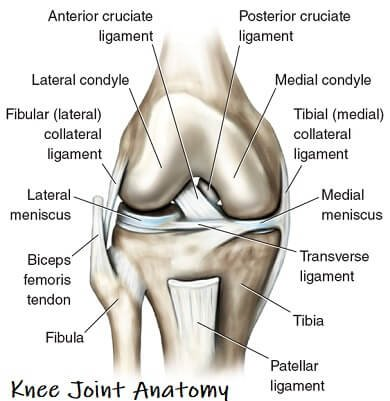
Patellar dislocation occurs when the kneecap (patella) moves out of its normal position in the groove at the front of the femur. This groove, called the trochlea, normally keeps the patella stable as you bend and straighten your knee. When the patella slips out—usually to the outer side—it disrupts normal movement and causes sudden pain, swelling, and a sense that the knee has “given way.”
A dislocation may be a single, isolated event, or it may recur if the knee has structural weaknesses or past injuries that have not fully healed. While many people recover well with proper treatment, ignoring symptoms may increase the risk of ongoing instability.
How Does Patellar Dislocation Impact Your Anatomy and Health?
When the kneecap dislocates, several structures can be damaged. Soft tissues such as ligaments and cartilage can stretch, tear, or bruise. In many cases, the medial patellofemoral ligament (MPFL) is injured because it normally holds the patella tightly in place. Damage to these tissues may cause long-term instability or difficulty with everyday activities, including walking, climbing stairs, kneeling, and squatting.
Cartilage damage is also a concern. When the patella moves out of its groove, the back of the kneecap and the groove can rub against each other, leading to early degeneration or osteoarthritis if left untreated. People may also experience weakness in the thigh muscles, particularly the quadriceps, because these muscles naturally tighten or compensate after injury. Over time, this may alter the knee's kinematics, increasing stress on surrounding joints and tissues.
Causes and Risk Factors for Patellar Dislocation
Patellar dislocation can affect anyone, but certain factors increase the likelihood of experiencing it:
- Age and activity: Teenagers and young adults involved in sports that require jumping, pivoting, or sudden changes of direction are at higher risk.
- Anatomical differences: Some people naturally have a shallow trochlear groove, higher-riding patella (patella alta), or misalignment of the leg bones that increases lateral pull on the kneecap.
- Previous dislocation: Once a dislocation has occurred, the risk of future episodes is significantly higher.
- Ligament laxity: People with looser ligaments or hypermobility conditions may experience instability more easily.
- Weakness in supporting muscles: Underdeveloped quadriceps or hip stabilisers make it harder for the patella to track properly.
- Gender: Females have a slightly higher risk due to natural differences in anatomy and ligament flexibility.
Symptoms of Patellar Dislocation
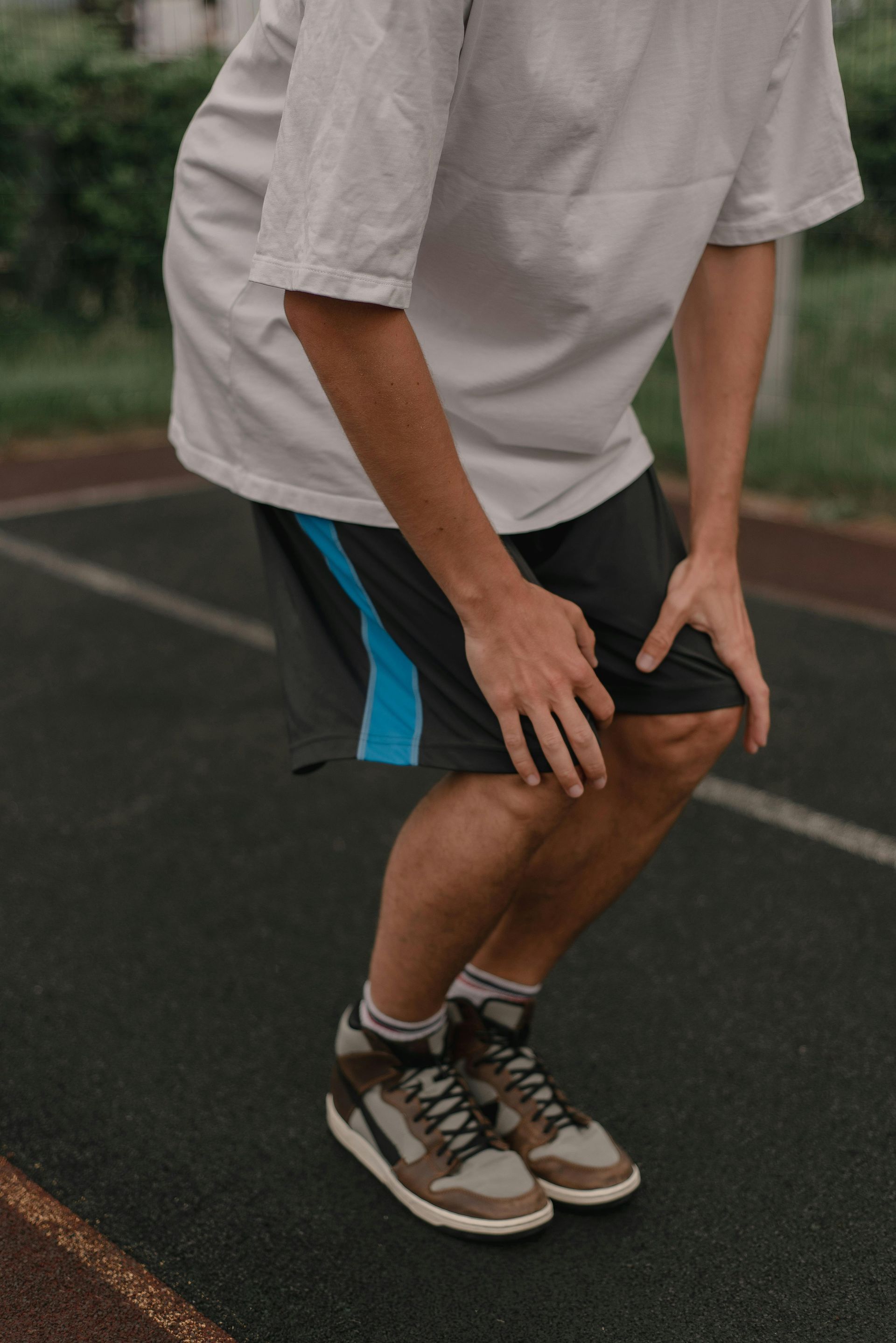
Symptoms of patellar dislocation are often immediate and noticeable. A person may feel or hear a pop, followed by strong pain at the front of the knee. Other symptoms include:
- Obvious deformity: The kneecap may appear out of place, usually toward the outer side.
- Sudden swelling: Fluid builds up quickly within the joint.
- Difficulty bending or straightening the knee: Movements become limited or painful.
- Instability or collapse: The knee may feel like it cannot bear weight or may buckle.
- Bruising and tenderness: Soft tissues around the joint may feel sore to the touch.
- Persistent discomfort after relocation: Even after the kneecap has returned to its position, pain and instability may persist.
Preventing Patellar Dislocation
Prevention focuses on building strength, improving knee alignment, and avoiding high-risk movements:
- Build quadriceps and hip strength:
Strong muscles help guide the patella smoothly through the trochlear groove.
- Improve flexibility: Stretching the hamstrings, quadriceps, and iliotibial band reduces uneven pulling forces.
- Correct foot mechanics: Orthotics or supportive footwear can improve leg alignment.
- Use proper technique in sports: Learning safe jumping, landing, and pivoting techniques reduces strain.
- Wear protective gear: Knee braces may help high-risk athletes maintain stability.
- Avoid overtraining: Fatigue increases coordination errors that may lead to injury.
Types of Patellar Dislocation
Patellar dislocations can be categorised based on mechanism, recurrence, and associated damage:
- Traumatic dislocation: Occurs due to a strong force such as a tackle or fall.
- Atraumatic dislocation: Happens without major force, often due to loose ligaments or anatomical differences.
- First-time dislocation: The first recorded episode of instability.
- Recurrent dislocation: Repeated episodes due to weakened structures or inadequate rehabilitation.
- Associated with fractures: Sometimes a piece of bone or cartilage breaks off when the patella dislocates.
Stages of Patellar Dislocation
Understanding the stages helps explain how the injury progresses:
- Stage 1: Initial injury: The kneecap is displaced, and soft tissues, such as the MPFL, may tear. Pain and swelling begin immediately.
- Stage 2: Relocation: The patella may spontaneously return to its natural position, or it may require manual reduction.
- Stage 3: Early healing: Swelling, bruising, and ligament damage heal slowly. Movement becomes restricted.
- Stage 4: Rehabilitation: Strengthening, stretching, and physiotherapy aim to restore normal motion and stability.
- Stage 5: Long-term outcome: If tissues heal well, stability returns. If not, there may be chronic instability or recurrent dislocations.
Diagnosis of Patellar Dislocation
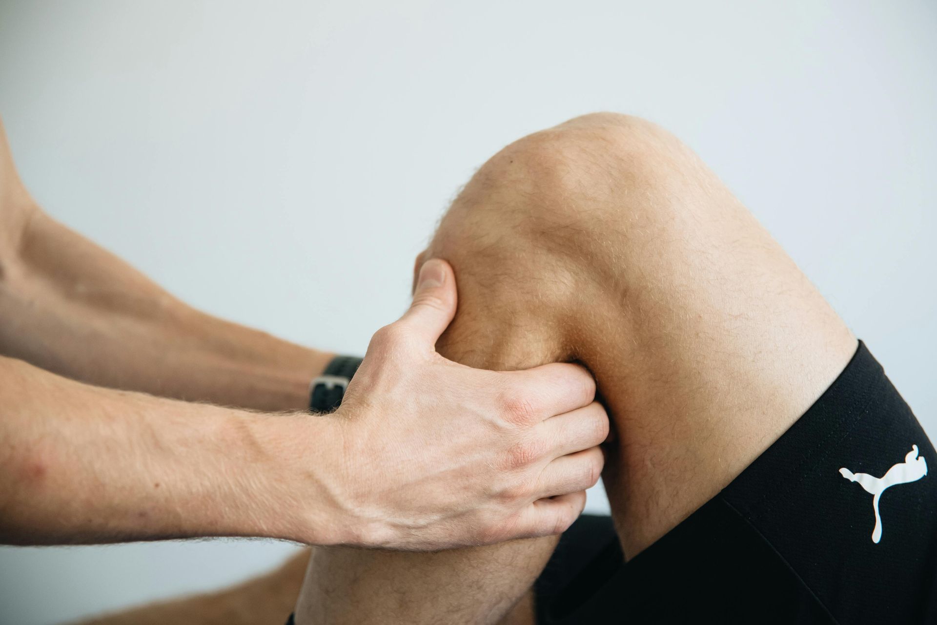
Diagnosis begins with a detailed history and physical examination. The doctor assesses swelling, deformity, and patellar movement. Special tests may be used to measure instability or ligament injury. Imaging helps confirm the diagnosis:
- X-ray: Identifies dislocation, misalignment, fractures, and bone shape differences.
- MRI: Shows ligament injuries, cartilage damage, and soft-tissue tears.
- CT scan: Provides detailed information on bone alignment, which is useful in certain complex cases.
Treatment for Patellar Dislocation
Treatment depends on the severity of the injury, presence of fractures, and risk of recurrence. Most first-time cases can be managed without surgery.
- Immediate care: Ice, rest, compression, elevation, and immobilisation reduce pain and swelling.
- Reduction of the patella: A trained clinician gently moves the kneecap back into position.
- Bracing: A stabilising brace helps maintain patellar alignment during early recovery.
- Physiotherapy: Strengthening the quadriceps and hip muscles improves patellar tracking. Stretching tight tissues restores normal movement.
- Pain management: Anti-inflammatory medications may reduce discomfort.
- Surgery: Considered if there is recurrent dislocation, severe ligament damage, loose bone fragments, or structural abnormalities. Procedures may include MPFL reconstruction, tibial tubercle realignment, or trochleoplasty.
What if Patellar Dislocation is Untreated?
Without proper treatment, patellar dislocation may lead to ongoing instability and repeated episodes of the kneecap slipping out. Recurrent dislocations can cause significant ligament damage, cartilage wear, and early arthritis. Chronic pain, reduced mobility, and decreased confidence in knee function are common long-term consequences. Everyday activities may become difficult, and sports participation may be limited. Early assessment and structured rehabilitation greatly reduce the risk of complications.